

Scientists found that messenger RNA (mRNA) molecules that carry genetic instructions to the far reaches of neurons in the brain tend to cluster together mostly because they are abundant, not because they move in coordinated groups.
The discovery, published in the Society for Neuroscience journal eNeuro, helps explain how neurons, which have some of the longest processes of any cells in the body, manage genetic instructions long distances from where they are made.
This fundamental process is critical to support neuronal communication and the modifications that occur at specific cellular communication sites called synapses along the neuron that are part of the cascade of molecular signals that occur during learning and the formation of memories.
When these processes break down — as they do in conditions such as Fragile X syndrome and some forms of autism — understanding the basic rules that guide RNA localization may help scientists pinpoint where things go wrong.
“When we compared every possible pair among the mRNAs we measured — 66 combinations in all — the simplest explanation fit best: Abundant mRNAs simply have more chances for their signals to overlap with the others we measured,” said Shannon Farris, assistant professor at Virginia Tech and its Fralin Biomedical Research Institute at VTC and senior author of the study. “It points to a flexible system in which mRNAs drift close together by chance. The real specificity seems to happen later at the synapse, where local signals decide how those genetic instructions are used.”
Some scientists have proposed that these mRNAs travel in distinct packets carrying specific combinations, while others have suggested that each message moves on its own. The new work leans toward the simpler idea that proximity mostly depends on how much of each message a cell produces.
Beyond providing a new look at basic biology, the insight helps clarify how neurons manage the messages that guide learning and memory, including processes that are disrupted in Fragile X syndrome, a genetic condition in which the protein that binds many of these RNAs is missing.
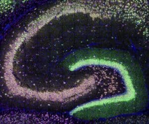
The team used single-molecule imaging in intact hippocampal tissue from wild-type mice to spatially map RNA messages, many of which are known to interact with a regulatory protein called FMRP. They measured the size and brightness of each glowing RNA dot and compared how often different RNAs appeared to spatially overlap.
Now that scientists have a clearer picture of how these molecules naturally arrange themselves, next steps include looking at how this system adapts during learning — and how it may go off-track in disorders that affect communication between neurons such as Fragile X syndrome.
The study was conducted by Farris, an assistant professor at the research institute and the Department of Biomedical Sciences and Pathobiology of the Virginia-Maryland College of Veterinary Medicine; first author Renesa Tarannum of the Virginia Tech Translational Biology, Medicine, and Health Graduate Program; Sharon Swanger, assistant professor at the research institute and the Department of Biomedical Sciences and Pathobiology; and Oswald Steward, director of the Reeve-Irvine Research Center for Spinal Cord Injury at the University of California, Irvine.
The research was supported by the National Institute of Neurological Disorders and Stroke, the National Institute of Mental Health, Virginia Tech’s Fralin Biomedical Research Institute, and the Brain and Behavior Research Foundation.
Original study: DOI 10.1523/ENEURO.0184-25.2025
By John Pastor